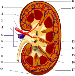
man kidneys viewed from behind with spine removed
The kidneys are paired organs, which have the production of urine as their primary function. Kidneys are seen in many types of animals, including vertebrates and some invertebrates. They are an essential part of the urinary system, but have several secondary functions concerned with homeostatic functions. These include the regulation of electrolytes, acid-base balance, and blood pressure. In producing urine, the kidneys excrete wastes such as urea and ammonium; the kidneys also are responsible for the reabsorption of glucose and amino acids. Finally, the kidneys are important in the production of hormones including vitamin D, renin and erythropoietin.
Located behind the abdominal cavity in the retroperitoneum, the kidneys receive blood from the paired renal arteries, and drain into the paired renal veins. Each kidney excretes urine into a ureter, itself a paired structure that empties into the urinary bladder.
Renal physiology is the study of kidney function, while nephrology is the medical specialty concerned with diseases of the kidney. Diseases of the kidney are diverse, but individuals with kidney disease frequently display characteristic clinical features. Common clinical presentations include the nephritic and nephrotic syndromes, acute kidney failure, chronic kidney disease, urinary tract infection, nephrolithiasis, and urinary tract obstruction.
The kidney has a bean-shaped structure, each kidney has concave and convex surfaces. The concave surface, the renal hilum, is the point at which the renal artery enters the organ, and the renal vein and ureter leave. The kidney is surrounded by tough fibrous tissue, the renal capsule, which is itself surrounded by perinephric fat, renal fascia (of Gerota) and paranephric fat. The anterior (front) border of these tissues is the peritoneum, while the posterior (rear) border is the transversalis fascia.
The substance, or parenchyma, of the kidney is divided into two major structures: superficial is the renal cortex and deep is the renal medulla. Grossly, these structures take the shape of 8 to 18 cone-shaped renal lobes, each containing renal cortex surrounding a portion of medulla called a renal pyramid (of Malphigi).[2] Between the renal pyramids are projections of cortex called renal columns (of Bertin). Nephrons, the urine-producing functional structures of the kidney, span the cortex and medulla. The initial filtering portion of a nephron is the renal corpuscle, located in the cortex, which is followed by a renal tubule that passes from the cortex deep into the medullary pyramids. Part of the renal cortex, a medullary ray is a collection of renal tubules that drain into a single collecting duct.
The tip, or papilla, of each pyramid empties urine into a minor calyx, minor calyces empty into major calyces, and major calyces empty into the renal pelvis, which becomes the ureter.
